


Breast Cancer: Screening and Diagnosis
October 10, 2023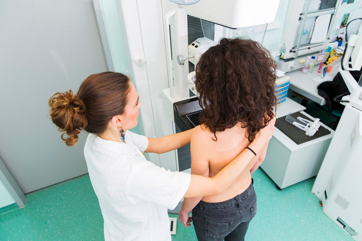
By: Val Enti
Categories: Cancer, Health & Wellness, Women's Health
Each year in the United States, approximately 264,000 women and 2,400 men are diagnosed with breast cancer. With one in 8 women affected, early detection is key to survival.
The guidelines regarding baseline mammograms for women with average risk of developing breast cancer has been updated. This article presents the recent mammogram update and provides a brief overview of the technologies used to detect breast cancer.
HANDS-ON BREAST EXAMINATIONS
Since the 1980s, more breast cancers are discovered through breast examinations than through any other screening method, according to a national database.
Clinical Breast Exam
In a clinical breast exam, a medical professional visually and manually checks a patient’s breasts for lumps, and skin or nipple abnormalities. Armpits are also examined.
The medical professional will also ask you about your age, when you began [or completed] menstruation, your personal and family breast health history, and your pregnancy history.
SCANS OR IMAGING
The most common scans associated with screening for breast cancer are ultrasounds and mammograms. For many women, these two imaging technologies provide answers and peace of mind. However, doctors may perform several additional tests or procedures to detect and confirm a cancer diagnosis.
- Breast Ultrasound
This device uses high-energy sound waves (echoes) that “bounce” off the area being examined. Ultrasounds are often used to take a closer look at an object found on a mammogram, and they are helpful with examining dense breasts. The images are able to determine if a breast lump is fluid-filled or solid. Typically, a fluid-filled lump is a breast cyst, which may not require treatment. A solid lump may or may not be cancerous. A lump may be followed over time using an ultrasound. Ultrasounds are also helpful when a specialist is performing a needle biopsy. Ultrasounds do not emit radiation.
- Mammogram
A mammogram is an X-ray of a woman’s breast that administers a low dose of radiation. It is usually taken from more than one angle to provide more information about any anomalies. These images provide different details about the breast than an ultrasound. Certain breast cancers do not show up on a mammogram, and it is more difficult to detect breast cancer using a mammogram if a woman has dense breasts. Women should be aware that false-positive and false-negative results can occur with a mammogram.
An important guideline change has occurred this year concerning when a woman should have her first mammogram. The U.S. Preventive Services Task Force recommends that women with average risk of developing cancer should now get their baseline mammogram at age 40, which has been lowered from age 50. The change is due to the fact that more younger women are presenting with breast cancer.
Breast Tomosynthesis
Breast tomosynthesis, also called 3-D mammography, is a relatively new technology. It takes images of the breast from many different angles and creates a three-dimensional picture of the tissue. This technology allows the breast specialist to examine dozens of images of the patient’s breast. Like a breast ultrasound, breast tomosynthesis may be particularly useful for women with dense breasts. However, as with a traditional mammogram, breast tomosynthesis can miss some breast cancers. The patient is exposed to radiation in this scan.
- Breast MRI or Magnetic Resonance Imaging
Women with high risk of developing breast cancer can benefit most from this technology. Detailed pictures of the breast are produced by radio waves combined with a powerful magnet and computing imaging. Fluids are injected into the patient for contrast. There is no radiation with this type of scan.
Research is constantly being conducted using new and experimental imaging techniques. Click here to learn more about these technologies.
BIOPSY
If imaging tests show a breast anomaly, the presence of cancer is confirmed through a biopsy that removes breast tissue for examination under a microscope. A biopsy can be performed through the use of a needle, sometimes together with imaging technologies.
A common biopsy procedure is an image-guided core needle biopsy — where the radiologist guides a needle into the tissue using imaging technology and then takes a sample for examination.
If the biopsy confirms the presence of cancer, it may be necessary to perform additional imaging or another biopsy to determine if the cancer has spread (metastasized) to the lymph nodes or other parts of the body.
Under certain circumstances, biopsies are also performed surgically.
The Bottom Line
About 13% of U.S. women will develop invasive breast cancer over the course of their lives, according to breastcancer.org. Knowing your risk factors is crucial. The bottom line is to advocate for your own health. Take charge of what is within your power to control, stay informed, and ask questions. It is your body and your health.
Learn more about Breast Care at Trinity Health Michigan or schedule your mammogram today.